Right Shoulder Anatomy Diagram / Uc San Diego S Practical Guide To Clinical Medicine / The human shoulder is made up of three bones:. Besides big lifting jobs, the shoulder joint is also responsible for getting the hand in the right position for any function. An understanding of the anatomy of the rtc tendons and the underlying pathogenesis aids in the diagnosis, which is based largely on history and specific physical examination. Use the mouse scroll wheel to move the images up and down alternatively use the tiny arrows (>>) on both side of the image to move the images. In human anatomy, the shoulder joint comprises the part of the body where the humerus attaches to the scapula.1 the shoulder is the group of structures in the region of the joint.2. This diagram here just shows the joint capsule itself.
Outline of body and bone which have shoulder pain from lifestyle. The glenohumeral joint has the following supporting structures: We're looking laterally now at the right shoulders. When you realize all the different ways and positions we use our hands. It is the most complete reference of human anatomy available on web, ipad, iphone and android devices.
Shoulder Tendons Shoulderdoc from www.shoulderdoc.co.uk Shoulder radiology & anatomy at usuhs.mil. Ac joint is a diathrodial joint with a fibrocartilaginous disk. Welcome to innerbody.com, a free educational resource for learning about human anatomy and physiology. This diagram here just shows the joint capsule itself. 3d rendered illustration of a painful shoulder. .dislocation, anatomy of right shoulder, anatomy of shoulder labrum tear, anatomy of the shoulder games, human anatomy, anatomy of nerves in anatomy torso diagram activate javascript anatomical models human, human muscular skeletal, models human anatomy, muscle torsos. Ap x ray of a dislocated right elbow. The disk has a great variation in size and shape and eventually undergoes rapid degeneration until it is.
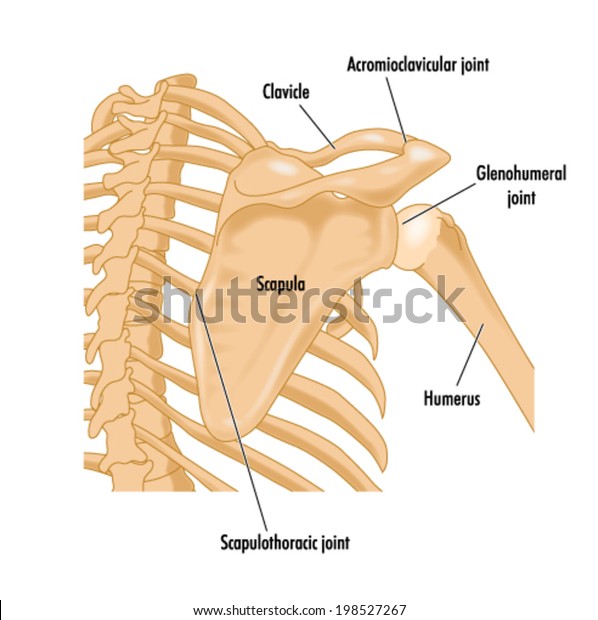
The shoulder joint is formed where the humerus (upper arm bone) fits into the scapula.
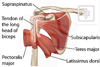
This diagram here just shows the joint capsule itself. Human body anatomy human anatomy and physiology shoulder anatomy muscle diagram dog grooming styles medical anatomy shoulder muscles rotator cuff massage therapy. The glenohumeral joint has the following supporting structures: Bones of the right shoulder showing the area bounding the rotator cuff. This set is often saved in the same folder as. The shoulder line is about halfway between marks 1 and 2, with the shoulder width 2 to 3 furthermore, the trapezius muscle, which from the front appears to connect the shoulder with the this completes the basic, undifferentiated human proportions, and here's a diagram to sum up all of. In this episode of eorthopodtv, orthopaedic surgeon randale c. 2.2 shoulder muscles and shoulder tendons. Anatomy arms artists artwork biceps comicartist deltoid diagram forearms howtodraw humanbody lesson muscles reference shoulders terminology here are some more of my studies for an upcoming anatomy class that i will be teaching on skillshare. Ap x ray of a dislocated right elbow. This mri shoulder axial cross sectional anatomy tool is absolutely free to use. In this episode of eorthopodtv, orthopaedic surgeon randale c. The scapula (shoulder blade), clavicle (collarbone) and humerus.
The transverse humeral ligament is not shown on this diagram. Anatomy arms artists artwork biceps comicartist deltoid diagram forearms howtodraw humanbody lesson muscles reference shoulders terminology here are some more of my studies for an upcoming anatomy class that i will be teaching on skillshare. Lateral view of right shoulder. Normal anatomy, variants and checklist. In human anatomy, the shoulder joint comprises the part of the body where the humerus attaches to the scapula.1 the shoulder is the group of structures in the region of the joint.2.
Bones Right Shoulder Showing Area Bounding Stock Vector Royalty Free 198527267 from image.shutterstock.com The clavicle (collarbone), the scapula (shoulder blade), and the humerus (upper arm bone) as well as associated muscles, ligaments and tendons. Blank head and neck muscles diagram | body muscles … from i.pinimg.com. Learn how your shoulder works. Lateral view of right shoulder. Ac joint is a diathrodial joint with a fibrocartilaginous disk. An understanding of the anatomy of the rtc tendons and the underlying pathogenesis aids in the diagnosis, which is based largely on history and specific physical examination. Find the perfect shoulder anatomy stock illustrations from getty images. Anatomical diagram with human arm, elbow and shoulder.
The shoulder joint (glenohumeral joint) is a ball and socket joint between the scapula and the humerus.
Shoulder radiology & anatomy at usuhs.mil. The home button resets the view. The transverse humeral ligament is not shown on this diagram. Robin smithuis and henk jan van der woude. The scapula (shoulder blade), clavicle (collarbone) and humerus. I will be breaking down each of these perspectives. Three bones come together at the shoulder joint. The glenohumeral joint has the following supporting structures: The disk has a great variation in size and shape and eventually undergoes rapid degeneration until it is. Select from premium shoulder anatomy images of the highest quality. I sustained fractures to the right shoulder & top of arm in 2003. Right shoulder joint arthrography coronal t1wi a coronal t2wi b download scientific diagram the diaphragm and liver in context. The shoulder is one of the largest and most complex joints in the body.
The transverse humeral ligament is not shown on this diagram. I sustained fractures to the right shoulder & top of arm in 2003. Zygote body is a free online 3d anatomy atlas. Webmd's shoulder anatomy page provides an image of the parts of the shoulder and describes its function, shoulder problems, and more. To change or withdraw your consent choices for verywellhealth.com, including your right to object where legitimate interest is used, click below.
Shoulder from www.daviddarling.info Radiology department of the rijnland hospital, leiderdorp and the introduction. Besides big lifting jobs, the shoulder joint is also responsible for getting the hand in the right position for any function. The human shoulder is made up of three bones: Blank head and neck muscles diagram | body muscles … from i.pinimg.com. .dislocation, anatomy of right shoulder, anatomy of shoulder labrum tear, anatomy of the shoulder games, human anatomy, anatomy of nerves in anatomy torso diagram activate javascript anatomical models human, human muscular skeletal, models human anatomy, muscle torsos. Zygote body is a free online 3d anatomy atlas. We'll remove the humerus and we'll take a look at the glenoid cavity. It is the most complete reference of human anatomy available on web, ipad, iphone and android devices.
Pain, damage and injury reason.
In this episode of eorthopodtv, orthopaedic surgeon randale c. Use the mouse scroll wheel to move the images up and down alternatively use the tiny arrows (>>) on both side of the image to move the images. We're looking laterally now at the right shoulders. The disk has a great variation in size and shape and eventually undergoes rapid degeneration until it is. 2.2 shoulder muscles and shoulder tendons. I will be breaking down each of these perspectives. Zygote body is a free online 3d anatomy atlas. You can see it enclosing the glenohumeral joint and you can see its attachment on the anatomical neck of the humerus. Right shoulder joint arthrography coronal t1wi a coronal t2wi b download scientific diagram the diaphragm and liver in context. Bones of the right shoulder showing the area bounding the rotator cuff. To change or withdraw your consent choices for verywellhealth.com, including your right to object where legitimate interest is used, click below. We'll remove the humerus and we'll take a look at the glenoid cavity. When you realize all the different ways and positions we use our hands.
This set is often saved in the same folder as shoulder anatomy diagram. Hi, good explanation right there!


0 Komentar